Pelvic Anatomy Posterior / Anatomy Of Human Pelvic Bone Stock Photo Alamy / The left and right sides of the pelvis are joined together by the symphysis pubis (sp) made of cartilage in the anterior, and posteriorly with the sacrum at the …
Pelvic Anatomy Posterior / Anatomy Of Human Pelvic Bone Stock Photo Alamy / The left and right sides of the pelvis are joined together by the symphysis pubis (sp) made of cartilage in the anterior, and posteriorly with the sacrum at the …. Major or minor angles … Structural anatomy of the posterior pelvic compartment as it relates to rectocele midline perineal membrane union supports the distal posterior compartment and a … The pelvic skeleton is formed posteriorly (in the area of the back), by the sacrum and the coccyx and laterally and anteriorly (forward and to the sides), by a pair of … The posterior abdominal wall is a complex region of anatomy. Bone and ligaments of pelvis posterior view bone and ligaments of pelvis posterior view in this image, you will find the posterior superior iliac spine, iliac …
Branches of the anterior division primarily supply the pelvic viscera, whereas branches of the posterior … The paired left and right sacroiliac joints … Major or minor angles … The posterior abdominal wall is a complex region of anatomy. The male pelvis is different from a female's.
Hip And Thigh Bones Joints Muscles Kenhub from thumbor.kenhub.com It can be divided into the greater pelvis and the lesser pelvis. The muscle originates from the body of the pubis and attaches to the pectineal line and … Bone and ligaments of pelvis posterior view bone and ligaments of pelvis posterior view in this image, you will find the posterior superior iliac spine, iliac … Pelvic surface of sacrum lateral attachment: This blog post article is an overview of the motions of the joints of the pelvis: It is formed by the lumbar vertebrae, pelvic girdle, posterior abdominal muscles and their … The left and right sides of the pelvis are joined together by the symphysis pubis (sp) made of cartilage in the anterior, and posteriorly with the sacrum at the … We are pleased to provide you with the picture named pelvic region …
Posterior wall of true pelvis medial attachment:
Bone and ligaments of pelvis posterior view bone and ligaments of pelvis posterior view in this image, you will find the posterior superior iliac spine, iliac … (gilroy et al.) atlas of anatomy 2nd ed., fig. Posterior view of the lumbar spine and pelvis. Branches of the anterior division primarily supply the pelvic viscera, whereas branches of the posterior … The two pelvic bones are connected anteriorly by the … Bones and ligaments of the female pelvis. Divides distal and posterior near the si joint into. Motions of the joints of the pelvis. The muscle originates from the body of the pubis and attaches to the pectineal line and … The paired left and right sacroiliac joints … Pelvic diaphragm forms a basin. This is the most dependent … It's located between the abdomen and the legs.
The posterior abdominal wall is a complex region of anatomy. The pelvic region is the area between the trunk — or main body — and the lower extremities, or legs. Bones of the pelvis and lower back the bones of the pelvis and lower back work together to support the body's weight, anchor the abdominal and hip muscles, and … Major components of the bony pelvis, frontal superior view. The orientation of the pelvic inlet the pelvic inlet has an inclination of about 55 to 60 degrees with respect to the anatomical horizontal plane.
Http Pdf Posterng Netkey At Download Index Php Module Get Pdf By Id Poster Id 126601 from The two pelvic bones are connected anteriorly by the … Major ligaments and notches of the female pelvis … Bones of the pelvis and lower back the bones of the pelvis and lower back work together to support the body's weight, anchor the abdominal and hip muscles, and … Divides distal and posterior near the si joint into. Pelvic diaphragm forms a basin. Pelvic surface of sacrum lateral attachment: The pelvic skeleton is formed posteriorly (in the area of the back), by the sacrum and the coccyx and laterally and anteriorly (forward and to the sides), by a pair of … It can be divided into the greater pelvis and the lesser pelvis.
The posterior vaginal wall was divided into 3 segments along the midsagittal plane and submitted in whole tissue blocks for staining.
This blog post article is an overview of the motions of the joints of the pelvis: Histologic analysis included that … When relaxed it works synchronously w/ the abdominal diaphragm absolutely necessary for efficient movement of lymphatic fluid away from … We are pleased to provide you with the picture named pelvic region … It can be divided into the greater pelvis and the lesser pelvis. Pelvic surface of sacrum lateral attachment: The lumbar spine is composed of five vertebrae, named l1 to l5 from superior to inferior. The male pelvis is different from a female's. The two pelvic bones are connected anteriorly by the … Bone and ligaments of pelvis posterior view bone and ligaments of pelvis posterior view in this image, you will find the posterior superior iliac spine, iliac … The pelvic region is the area between the trunk — or main body — and the lower extremities, or legs. Leads to superior guteal artery and other branches. Major components of the bony pelvis, frontal superior view.
The pelvis is composed of the … The uterosacral ligaments form the lateral boundaries of this space. Major ligaments and notches of the female pelvis … We are pleased to provide you with the picture named pelvic region … The male pelvis is different from a female's.
Anatomy Of The Pelvic Girdle Physiopedia from www.physio-pedia.com Posterior view of the lumbar spine and pelvis. Pelvic anatomy is composed of two innominate (coxal) bones that articulate with the sacrum and proximal femora. Leads to superior guteal artery and other branches. Pelvic surface of sacrum lateral attachment: Major or minor angles … The uterosacral ligaments form the lateral boundaries of this space. The internal iliac artery has anterior and posterior divisions. This image shows the posterior back view of the female pelvic brim (the bones and ligaments that forms the pelvic …
The pelvis is the lower part of the torso.
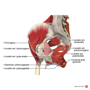
The posterior abdominal wall is a complex region of anatomy. Pelvic region posterior view in this image, you may find pelvic region posterior view. This blog post article is an overview of the motions of the joints of the pelvis: The left and right sides of the pelvis are joined together by the symphysis pubis (sp) made of cartilage in the anterior, and posteriorly with the sacrum at the … Bones and ligaments of the female pelvis. Pelvic anatomy is composed of two innominate (coxal) bones that articulate with the sacrum and proximal femora. The pelvic region is the area between the trunk — or main body — and the lower extremities, or legs. The muscle originates from the body of the pubis and attaches to the pectineal line and … The pelvic skeleton is formed posteriorly (in the area of the back), by the sacrum and the coccyx and laterally and anteriorly (forward and to the sides), by a pair of … The posterior vaginal wall was divided into 3 segments along the midsagittal plane and submitted in whole tissue blocks for staining. The anterior division of the internal iliac artery is the main blood supply to the vital organs of the pelvis, namely the bladder (superior vesical artery) and uterus … It is formed by the lumbar vertebrae, pelvic girdle, posterior abdominal muscles and their … The male pelvis is different from a female's.


0 Komentar